


山本 博章
(やまもと・ひろあき)
Hiroaki Yamamoto
略歴
- 東北大学大学院理学研究科博士課程後期修了
- 東北大学教養部生物学科助手、同理学部生物学科助教授、同大学院生命科学研究科助教授、准教授を経て本学へ
色素細胞の分化と機能を制御する分子機構
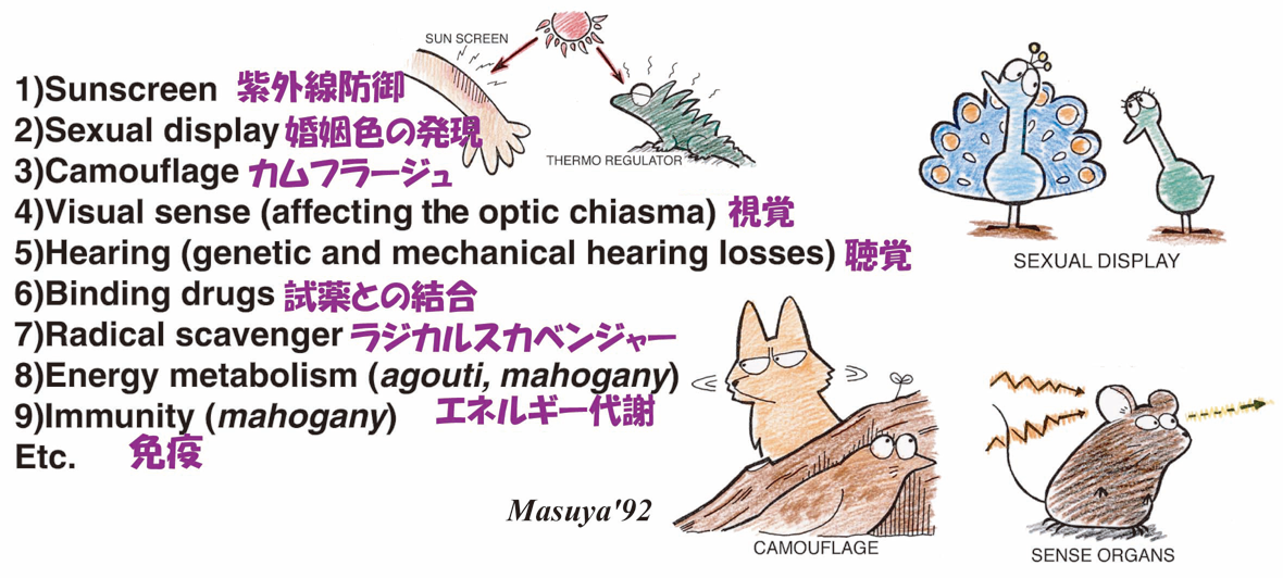
この細胞の機能については、紫外線防御やカムフラージュ、また婚姻色の発現等に関わることがよく知られるところですが、それだけでなく、多くの機能を持っています。たとえば、我々ヒトが発生させることができる色素細胞はメラニン合成をおこなうメラニン色素細胞だけですが(魚類や両生類また爬虫類はメラニン色素以外の色素も合成することができます)、これら色素細胞は視聴覚にも必須であることが分かっています。このメラニン色素細胞の発生やメラニン合成に深くかかわる遺伝子にMitf (microphthalmia associated transcription factor) と呼ばれる転写因子をコードする遺伝子が関わっています。
これまでのところ私たちが持つ約20,000あまりの遺伝子(座)の中で400遺伝子(座)近くが何らかのかたちで色の発現にかかわっていることが分かっています。この中で「すべての」メラニン色素細胞の「発生」に必須であるのは、その多くの突然変異遺伝子(対立遺伝子)の解析から、このMitf以外まだ知られていません。従いまして、メラニン色素細胞の発生や機能の発現解析には、この遺伝子の働きを調べることが必須となります。
最近分かったこと
1、Mitfは色素細胞の発生だけでなく、皮膚に分布する色素細胞の形や細胞内の色素顆粒(メラノソームと呼ばれます)の移送にもかかわる(Kawasaki eetal.,2008, 2011)。
2、Mitfは、発生中の脳から形成されその活性が視覚に必須である網膜色素上皮と呼ばれるメラニン合成細胞の一領域で細胞分裂を抑える働きをしている(Tsukiji etal., 2009)。
3、Mitfのある分子型を欠損して黒眼で白毛色となるハツカネズミ突然変異体は難聴であるが、この変異体を用いて、聴覚に必須の内耳色素細胞では酸化ストレス耐性に重要な機能を持つタンパク質をコードする遺伝子が特異的に強く発現していることを発見した(Uehara etal., 2009)。
4、上記マウス変異体の眼の外側を覆う脈絡膜に定着した色素細胞が、当該部位に発達する脈管系の早い血流速度を保障している可能性を発見した(Shibuya etal., accepted 2018)。
これらのことも踏まえて、私たちのグループは、色素細胞はすべてのストレスを吸収し緩和する細胞として進化してきたのではないかと、想像をたくましくしています。従いまして、この色素細胞がこれまで環境にどう反応してきたのか、またこれからどのように機能分化してゆくのか、大変興味深いと思っているところです。これらを解析することによって、私たちがどのように環境ストレスを感じまたそれに応答しているのかをモニターできる一モデル系を確立できるだけでなく、さらには積極的なストレス緩和システムの構築につなげることができるのではないかと期待しています。
図1:色素細胞の機能(や関わる遺伝子の多面発現)
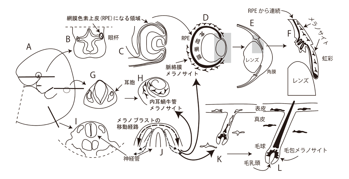
図2:哺乳動物の色素細胞系譜: Mammalian pigment cell lineages発生中の脳に形成される眼杯からは網膜色素上皮(RPE)が分化する。胚の背側に形成される脊椎動物特異的な神経冠(堤)に由来するメラニン色素細胞(メラノサイト)の前駆細胞(メラノブラスト)が、耳胞の領域を含め全身に移動する様子を示す。
図3:黒眼白毛色マウス突然変異体(Mitf mi-bw/Mitf mi-bw): A black-eyed white mouse mutant このマウスは網膜色素上皮は正常に発生するので黒眼となるが、脊椎動物特的な神経冠(堤)由来の色素細胞が発生できず、白毛色で難聴を示す。

- 研究の応用領域
- 化粧品、医薬品、診断薬の開発
- 産官学連携で求めるパートナー
- 色素細胞が関与する現象に関わる基礎・応用・臨床研究者。化粧品・医薬品開発の関連企業、大学、国・地方自治体の研究機関
MOLECULAR MECHANISMS CONTROLLING PIGMENT CELL DIFFERENTIATION AND FUNCTION
One of the long-term goals of our research group is to elucidate the molecular mechanisms by which pigment cells differentiate and function in multicellular organisms in order to infer the evolution of those mechanisms and to predict the roles of these cells. To this end, phylogenetic analyses of these mechanisms and surveying uncovered roles of these cells are a central and indispensable line of our research.
Why pigment cells?
1) Ease of applying developmental genetics. In mice, nearly 400 loci are known that affect pigmentation. Those features allow us to analyze the molecular mechanisms by which these cells differentiate to pigment cells.
2) Mutations are easily identified by visible changes of the coat (skin) colors.
3) Mutations usually do not critically affect fertility or survival.
Roles of pigment cells: 1) Sun screen, 2) sexual display, 3) camouflage, 4) visual and auditory senses, 5) Binding drugs, 6) Radical scavenger, 7) Energy metabolism, 8) Immunity, etc.
Our recent findings:
1) The transcription factor Mitf-M (M isoform) is essential for regulating the expression of the skin (coat) color (Yajima et al., 1999)
2) Isolation and characterization of ascidian Mitf: our data support the concept that the acquisition of multiple promoters (isoforms) by an ancestral Mitf gene has allowed the evolution of multiple pigment cell types (Yajima et al., 2003)
3) Mitf activity is critically regulated during the normal development of pigment cells: over-expression of either normal or a dominant-negative form of Xenopus Mitf induces the microphthalmia phenotypes (Kumasaka et al., 2005).
4) Mitf is involved in the regulation of melanosome transport and the level of dendricity in melanophores (Kawasaki et al., 2008).
5) Cochlear melanocytes in the stria vascularis (but not in hair follicles) specifically express Gsta4 which is deeply involved in anti-stress responses (Uehara et al., 2009).
6) Mitf uniquely regulates both differentiation and cell proliferation in the developing RPE by regulating the expression of p27kip1, one of the cyclin-dependent kinase inhibitors (Tsukiji et al., 2009).
7) ET3/Ednrb2 signaling is critically involved in regulating melanophore migration in Xenopus (Kawasaki-Nishihara et al., 2011)
8) Otx2 is deeply involved in the regional specification of the developing RPE (Nishihara et al., 2012)
Ongoing projects:
Analyses of
・Melanocyte (melanophore) migration
・RPE development
・Hair cycle (a biological clock with long-span periodic activity)
・Function of melanocytes and RPE
Your suggestions and collaboration would be very much appreciated
Shibuya, H., Watanabe, R., Maeno, A., Ichimura, K., Tamura, M., Wakana, S., Shiroishi, T., Ohba, K., Takeda, K., Tomita, H., Shibahara, S. and Yamamoto, H. Melanocytes contribute to the vasculature of the choroid. Genes & Genetic Systems, accepted (2018).
Tabata, H., Koinui, A., Ogura, A., Nishihara, D. and Yamamoto, H. A novel nuclear localization signal spans the linker of the two DNA-binding subdomains in the conserved paired domain of Pax6. Genes & Genetic Systems, accepted (2018).
Nishihara, D., Yajima, I., Tabata, H., Nakai, M., Tsukiji, N., Katahira, T., Takeda, K., Shibahara, S., Nakamura, H and Yamamoto, H. Otx2 is involved in the regional specif ication of the developing retinal pigment epithelium by preventing the expression of Sox2 and Fgf8, factors that induce neural retina dif f erentiation. PloS ONE, 7(11), e48879. (2012)
Kawasaki-Nishihara, A., Nishihara, D., Nakamura, H. and Yamamoto, H. ET3/Ednrb2 signaling is critically involved in regulating melanophore migration in Xenopus. Developmental Dynamics, 240, 1454-1466. (2011)
Tsukiji, N., Nishihara, D., Yajima, I., Takeda, K., Shibahara, S. and Yamamoto, H. Mitf functions as an in ovo regulator for cell dif f erentiation and proliferation during development of the chick RPE. Developmental Biology 326, 335-346. (2009)